

Summary of Product Characteristics (SmPC) - Tauvid 1 900 MBq/ml solution for injection
1 NAME OF THE MEDICINAL PRODUCT
Tauvid 1 900 MBq/mL solution for injection
2 QUALITATIVE AND QUANTITATIVE COMPOSITION
Tauvid 1 900 MBq/mL solution for injection
Each mL of solution for injection contains 1 900 MBq of flortaucipir (18F) at the date and time of calibration (ToC).
The activity per vial ranges from 1 900 MBq to 28 500 MBq at the ToC.
Fluorine (18F) decays to stable oxygen (18O) with a half-life of approximately 110 minutes by emitting a positron radiation of 634 keV, followed by photonic annihilation radiation of 511 keV.
Excipient(s) with known effect
Each mL of solution contains up to 79 mg of ethanol, 3.4 mg of sodium. For the full list of excipients, see section 6.1.
3 PHARMACEUTICAL FORM
Solution for injection (injection).
Clear, colourless solution.
4 CLINICAL PARTICULARS
4.1 Therapeutic indications
This medicinal product is for diagnostic use only.
Flortaucipir (18F) is a radiopharmaceutical indicated for Positron Emission Tomography (PET) imaging of the brain to estimate the density and distribution of the aggregated tau neurofibrillary tangles (NFTs) of Alzheimer’s disease (AD) in adult patients with cognitive impairment who are being evaluated for AD. Flortaucipir (18F) is an adjunct to clinical and other diagnostic evaluations.
4.2 Posology and method of administration
A PET scan with flortaucipir (18F) should be requested by physicians skilled in the clinical management of neurodegenerative disorders.
Tauvid images should only be interpreted by readers trained in the interpretation of PET images with flortaucipir (18F).
Posology
The recommended single intravenous dose for flortaucipir (18F) injection is 370 MBq (10 mCi) of
flortaucipir (18F) in a dose volume of ≤10 mL.
Special populations
Elderly population
No dose adjustment is recommended based on age.
Renal and hepatic impairment
Careful consideration of the activity to be administered is required since an increased radiation exposure is possible in these patients (see section 4.4).
Paediatric population
There is no relevant use of flortaucipir (18F) in the paediatric population.
Method of administration
Flortaucipir (18F) is for intravenous use and multidose use.
The radiopharmaceutical dose should be measured by a suitable radioactivity measurement system and inspected for particulate matter or discolouration prior to administration.
The dose is administered by intravenous bolus injection, followed by a flush of sodium chloride 9 mg/mL (0.9 %) solution for injection to ensure full delivery of the dose. Flortaucipir (18F) may be diluted up to 1:5 based on volume, with sodium chloride 9 mg/mL (0.9 %) solution for injection.
Only authorised persons qualified by training and experience should receive, use and administer flortaucipir (18F). Use appropriate safety measures to minimize unnecessary radiation exposure to clinical personnel, patients, and the public.
Image acquisition
A 20 minute PET image should be acquired starting approximately 80 minutes after intravenous injection of flortaucipir (18F). Patients should be supine with the head positioned to centre the brain, including the cerebellum, in the PET scanner field of view. Reducing head movement with tape or other flexible head restraints may be employed.
4.3 Contraindications
Hypersensitivity to the active substance or to any of the excipients listed in section 6.1.
4.4 Special warnings and precautions for use
Individual benefit/risk justification
For each patient, the radiation exposure must be justifiable by the likely benefit. The activity administered should in every case be as low as reasonably achievable to obtain the required diagnostic information.
Limitations of use
Flortaucipir (18F) binds selectively to AD-type neurofibrillary tangles. The safety and effectiveness of flortaucipir (18F) to assess the distribution of tau resulting from chronic traumatic encephalopathy (CTE), non-AD type dementias, or neurodegenerative conditions have not been established.
Renal and hepatic impairment
Flortaucipir (18F) is excreted through the hepatobiliary and renal systems. Patients with hepatic or renal impairment have the potential of increased radiation exposure (see section 4.2). Careful consideration of the benefit risk ratio in these patients is required since an increased radiation exposure is possible.
Interpretation of flortaucipir (18F) images
The goal of the read is to identify and locate areas of flortaucipir activity in the neocortex that are greater than the background activity (background activity is defined as up to 1.65-fold the measured cerebellar average). For optimal display, select a colour scale with a rapid transition between two distinct colours and adjust the scale so that the transition occurs at the 1.65-fold threshold. Examine the posterolateral temporal (PLT), occipital, parietal, and frontal regions bilaterally. Neocortical activity in either hemisphere contributes to image interpretation. Activity in white matter or regions outside the brain does not contribute to image interpretation. To help identify the PLT region, consider subdividing the temporal lobe into four quadrants as instructed below. Activity in the anterior and medial temporal lobe does not contribute to image interpretation of an AD flortaucipir (18F) pattern.
Image display and orientation
Display images in the transverse, sagittal, and coronal planes. Reorient images to remove head tilt in the transverse and coronal plane. Use a sagittal slice just off the midline to align the inferior frontal and inferior occipital poles in the horizontal plane.
Select and adjust the colour scale
To create a visual threshold for positivity:
- Draw a region of interest around the cerebellum in the transverse plane.
- Select the plane to go through the cerebellum at the maximum cross-sectional area of the cerebellum.
- Record the mean activity or cerebellar counts (MCC). The region of interest should be drawn with the scan in grey scale and in the transverse plane as seen in the example in Figure 1.
Figure 1: Example of Cerebellar Region of Interest 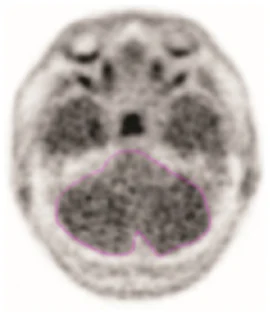
- Select a colour scale for image display that has a rapid transition between two distinct colours in the general range of 25 % to 60 % of maximum intensity.
- Set the upper contrast value (UCV) of the colour scale. Use the following formula to set the visual threshold of 1.65 x MCC to match the rapid transition in the colour scale:
UCV = (MCC x 1.65) x (100 % / % level of colour transition)
Preparation for image interpretation
- Before interpreting the image, review the brain to determine the lobar anatomy. Interpret the images by first evaluating the temporal lobes, followed by occipital, parietal, and frontal lobes bilaterally.
- To evaluate the temporal lobes, subdivide them into four quadrants by placing the horizontal crosshair immediately posterior to the brainstem nuclei and then scrolling inferiorly to place the vertical crosshair through the widest portion of the temporal pole, thus obtaining the anterolateral temporal (ALT), anterior mesial temporal (AMT), posterolateral temporal (PLT) and posterior mesial temporal (PMT) quadrants. See Figure 2 for an example (the left and right image panels show the same scan in two different colour scales).
Figure 2: Temporal Lobe Quadrants
Colour Scale 1 Colour Scale 2
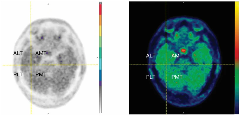
Image interpretation errors can occur with flortaucipir (18F) imaging. Interpret the PET flortaucipir (18F) images based upon the pattern and density of the radioactive signal within the neocortical grey matter (not within white matter or in regions outside of the brain). Only uptake of tracer in the neocortical grey matter regions should contribute to scan interpretation.
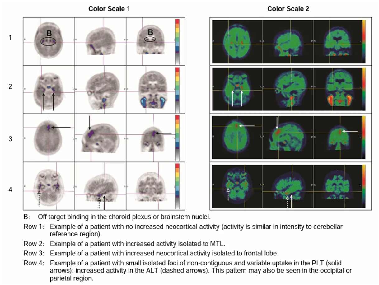
Off-target binding may be seen in the choroid plexus, striatum, and brainstem nuclei. Small foci of non-contiguous tracer uptake may lead to false positive interpretation. Interpret scans that have isolated or non-contiguous, small foci in any region with caution. Some scans may be difficult to interpret due to image noise or motion artifact. For cases where there is uncertainty as to the location of neocortical uptake, use co-registered anatomical imaging to improve localisation of uptake.
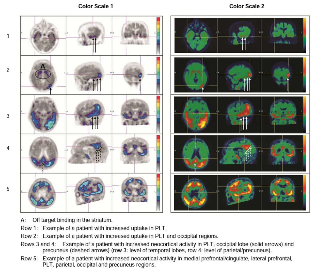
Flortaucipir (18F) activity patterns supportive of AD diagnosis: Scans with increased neocortical activity in the posterolateral temporal (PLT), occipital, or parietal/precuneus region(s), with or without frontal region(s) activity, indicate the presence of tau B3 NFTs (tau pathology scoring; section 5.1). In patients with an appropriate clinical workup, a scan indicating the presence of B3 NFTs is supportive of an AD diagnosis. Neocortical activity in either hemisphere can contribute to identification of the pattern.
AD flortaucipir (18F) patterns fall into one of two categories:
- Moderate AD flortaucipir (18F) pattern (Figure 3, rows 1 and 2): increased neocortical activity in the PLT or occipital region(s).
- Advanced AD flortaucipir (18F) pattern (Figure 3, rows 3, 4, and 5): increased neocortical activity in the parietal/precuneus region(s), or increased activity in the frontal region(s) accompanied by increases in the PLT, parietal, or occipital region(s).
Figure 3. AD Diagnostic Scan Examples
Negative flortaucipir (18F) pattern:
Scans without increased neocortical activity, or with increased neocortical activity isolated to the mesial temporal, anterolateral temporal, and/or frontal regions represent negative flortaucipir (18F) patterns (Figure 4). A negative flortaucipir (18F) scan pattern is insufficient to conclude the absence of AD neuropathology. A negative flortaucipir (18F) pattern does not rule out the presence of B2 or lower NFT AD neuropathology (see section 5.1).
Figure 4. Negative Scan Examples
After the procedure
Close contact with infants and pregnant women should be restricted during the initial 4 hours following the injection.
Sodium
Tauvid 1 900 MBq/mL solution for injection contains up to 34 mg sodium per dose, equivalent to less than 2 % of the WHO recommended maximum daily intake of 2 g sodium for an adult.
Ethanol
This medicinal product contains 790 mg of alcohol (ethanol) in each 10 mL dose, which is equivalent to 11.3 mg/kg (administered to an adult with 70 kg). The amount in 10 mL of this medicinal product is equivalent to less than 20 mL beer or 8 mL wine. The small amount of alcohol in this medicinal product will not have any noticeable effects.
4.5 Interaction with other medicinal products and other forms of interaction
Potential for flortaucipir (18F) to affect other drugs
In vitro, flortaucipir (19F) (the non-radioactive form of flortaucipir (18F)) did not cause clinically relevant inhibition of the activity of several cytochrome P450 enzymes (CYPs 3A, 1A2, 2B6, 2C8, 2C9, 2C19, and CYP2D6) or the transporter P-glycoprotein (Pgp).
Potential for other drugs to affect flortaucipir (18F)
In vitro, flortaucipir (19F) is a substrate of CYP1A2 (primarily) and CYP2D6 (minor contribution).
However, in vitro studies suggest CYPs play a minor role in the overall clearance of flortaucipir compared to other clearance routes such as aldehyde oxidase. Therefore, inhibitors of CYP1A2 and CYP2D6 are unlikely to cause clinically meaningful changes in the pharmacokinetics of flortaucipir (18F).
4.6 Fertility, pregnancy and lactation
Women of childbearing potential
When an administration of radiopharmaceuticals to a woman of childbearing potential is intended, it is important to determine whether or not she is pregnant. Any woman who has missed a period should be assumed to be pregnant until proven otherwise. If in doubt about her potential pregnancy (if the woman has missed a period, if the period is very irregular, etc.), alternative techniques not using ionising radiation (if there are any) should be offered to the patient. It is recommended that women of reproductive potential, unless using effective birth control, should abstain from intercourse for 24 hours (> 10 half-lives of radioactive decay for the 18F isotope) following flortaucipir (18F) administration.
Pregnancy
Radionuclide procedures carried out on pregnant women also involve radiation dose to the foetus. Any radiopharmaceutical, including flortaucipir (18F), has a potential to cause foetal harm. Use of flortaucipir (18F) is not recommended in pregnant women. Only imperative investigations should therefore be carried out during pregnancy, when the likely benefit far exceeds the risk incurred by the mother and foetus. There are no available data on flortaucipir (18F) use in pregnant women. No animal reproduction studies have been conducted with flortaucipir (18F).
Breast-feeding
Lactation studies have not been conducted in animals. It is not known if flortaucipir (18F) is excreted in human milk. Before administering radiopharmaceuticals to a mother who is breastfeeding, consideration should be given to the possibility of delaying the administration of radionuclide until the mother has ceased breastfeeding, and to what is the most appropriate choice of radiopharmaceuticals, bearing in mind the secretion of activity in breast milk. If the administration is considered necessary, breastfeeding should be interrupted for 24 hours and the expressed feeds discarded.
Close contact with infants should be restricted during the initial 4 hours following injection.
Fertility
It is not known if flortaucipir (18F) has any effect on fertility.
4.7 Effects on ability to drive and use machines
Tauvid has no or negligible influence on the ability to drive and use machines.
4.8 Undesirable effects
Summary of the safety profile
The safety profile of flortaucipir (18F) is based on its administrations to 4 652 subjects in clinical trials.
Tabulated list of adverse reactions
Frequencies are defined as uncommon (≥ 1/1 000 to < 1/100). While they may in reality occur at lower frequencies than indicated below, the size of the source database did not allow for the assignment of frequency categories lower than the category “uncommon” (≥ 1/1 000 to <1/100).
| System organ class | Adverse reaction and frequency |
| Nervous system disorders | Uncommon: headache Uncommon: dysgeusia |
| General disorders and administration site conditions | Uncommon: injection site pain |
| Investigations | Uncommon: blood pressure increaseda |
aIncludes hypertension, blood pressure systolic increased, and hypertensive urgency
Exposure to ionising radiation is linked with cancer induction and a potential for development of hereditary defects. As the effective dose is 8.7 mSv when the maximal recommended activity of 370 MBq is administered, these adverse reactions are expected to occur with a low probability.
Reporting of suspected adverse reactions
Reporting suspected adverse reactions after authorisation of the medicinal product is important. It allows continued monitoring of the benefit/risk balance of the medicinal product. Healthcare professionals are asked to report any suspected adverse reactions via the Yellow Card Scheme, website: www.mhra.gov.uk/yellowcard or search for MHRA Yellow Card in the Google Play or Apple App Store.
4.9 Overdose
Due to the small quantity of flortaucipir (18F) in each dose, overdose is not expected to result in pharmacological effects. In the event of administration of a radiation overdose, the absorbed dose to the patient should be reduced where possible by increasing the elimination of the radionuclide from the body by frequent micturition and defaecation. It might be helpful to estimate the effective dose that was applied.
5 PHARMACOLOGICAL PROPERTIES
5.1 Pharmacodynamic properties
Pharmacotherapeutic group: diagnostic radiopharmaceutical, central nervous system, ATC code: V09AX07
Mechanism of action
Flortaucipir (18F) binds to aggregated tau protein. In brains of patients with AD, paired helical filament (PHF) tau forms aggregates that combine to form neurofibrillary tangles (NFTs), a required component of the neuropathological diagnosis of AD. In vitro, flortaucipir (18F) binds to PHF tau purified from brain homogenates of donors with AD. Weak binding and poor colocalisation was observed for tau aggregates from other non-AD tauopathies. In vivo, flortaucipir (18F) is differentially retained in neocortical areas that contain aggregated tau.
Pharmacodynamic effects
At the chemical concentrations used for diagnostic examinations, flortaucipir (18F) does not appear to have any pharmacological activity.
In studies that included cognitively normal and cognitively impaired patients, after intravenous administration, scans starting at approximately 80 minutes after injection and lasting for 20 minutes had the highest test-retest reproducibility.
Clinical efficacy and safety
The safety and diagnostic performance of flortaucipir (18F) imaging to estimate the density and distribution of aggregated tau neurofibrillary tangles (NFTs), compared to post-mortem examination, was evaluated in 2 pivotal AD neuropathologic correlation clinical studies. In each study, 5 independent readers, who were blinded to clinical information, interpreted flortaucipir (18F) imaging as positive or negative. Independent pathologists, who were blinded to clinical and imaging results, subsequently conducted post-mortem examination of the brains. The pathologists recorded tau NFT scores derived from Braak staging (ranging from B0 to B3) (Table 1).
Table 1: Tau Pathology Scoring
| Tau Pathology Score | Distribution of Tau NFTs in the Brain |
| B0 | No NFTs |
| B1 | NFTs limited to transentorhinal brain region |
| B2 | B1 + NFTs limited to limbic brain regions |
| B3 | B2 + NFTs distributed throughout the neocortex |
In Study 1, reader interpretation of pre-mortem flortaucipir (18F) scans from 64 terminally ill patients was compared to the findings from post-mortem brain examinations. Of the 64 patients, the mean age was 83 years (range 55 to 100); 34 were female; 49 had dementia, 1 had mild cognitive impairment, and 14 had no cognitive impairment on clinical evaluation around the time of flortaucipir (18F) imaging. The study evaluated the performance of AD flortaucipir (18F) pattern scans for distinguishing B3 (positive) from B0-B2 (negative) tau pathology.
Study 2 evaluated diagnostic performance in 82 terminally ill patients (the same 64 patients from Study 1, plus 18 additional terminally ill patients) and reader performance in scans from the terminally ill patients plus 159 patients with cognitive impairment being evaluated for AD.
The diagnostic performance of an AD flortaucipir (18F) pattern to confirm the presence of AD neuropathology from both studies is presented in Table 2.
Table 2: Diagnostic performance of flortaucipir (18F) scan among autopsied patients - Studies 1 and 2
| Truth Standard | Study (N) | Sensitivity (%) (median and range) | Specificity (%) (median and range) |
B3 NFT (Primary Analysis 1) | Study 1 (64) | 92 (92 – 100) | 76 (52 – 92) |
Study 2 (82) | 89 (87 – 94) | 77 (63 – 91) |
An exploratory analysis using quantitation of 60 flortaucipir (18F) PET scans with autopsy truth standard from Study 1 was performed using an AD-signature weighted neocortical target volume of interest and an individual white matter-based reference region. A standardized uptake value ratio (SUVr) cutoff of 1.113 was determined by the Youden index method. This cutoff correctly identified the presence of B3 NFT in 84.2 % (32/38) of the positive truth standard cases and was 100 % (22/22) accurate to identify cases with B2 NFT score or lower.
Reader Performance
In both studies there was substantial to excellent reader agreement. In Study 1, for all cases with a visual read (whether or not the patient came to autopsy; n=105), Fleiss’ kappa for inter-reader agreement across the 5 readers was 0.80 (95 % confidence interval 0.74 to 0.86). In Study 2, the Fleiss’ kappa for all cases read including both scans from Study 1 autopsy subjects and scans from subjects with clinically defined MCI and AD (n=241) was 0.87 (95 % confidence interval 0.83 to 0.91).
Paediatric population
The licensing authority has waived the obligation to submit the results of studies with Tauvid in all subsets of the paediatric population as the disease or condition for which the specific medicinal product is intended only occurs in adults (see section 4.2 for information on paediatric use).
5.2 Pharmacokinetic properties
Distribution
Flortaucipir (18F) is distributed and metabolised rapidly throughout the body. Less than 10 % of the injected (18F) radioactivity remains in blood 5 minutes after administration, and less than 5 % is present 10 minutes after administration.
Organ uptake
Maximum brain uptake of radioactivity occurs within several minutes of injection, followed by rapid brain clearance during the first 45 minutes following injection.
Healthy controls show relatively low levels of flortaucipir (18F) retention in the cortex and cerebellum. In amyloid-positive AD and MCI subjects, cortical regions show significantly greater uptake compared with cognitively normal controls. In AD and MCI subjects, as in controls, there is low retention in the cerebellum. In older controls who are amyloid negative, high retention in the choroid plexus, striatum, and brainstem nuclei has been observed and binding in these regions is presumed to be off-target.
Elimination
The residual 18F in circulation during the 80 to 100 minute imaging window is composed of approximately 28 %-34 % parent drug with the remainder being metabolites.
Clearance occurs primarily by hepatobiliary and renal excretion.
Half-life
Flortaucipir is very rapidly cleared from circulation post-intravenous injection. Plasma radioactivity (including parent flortaucipir and all its metabolites) is below 10% of the theoretical maximum concentration by 5 minutes post-dose. The radioactive half-life of 18F is 110 minutes.
Renal/ Hepatic impairment
The pharmacokinetics in patients with renal or hepatic impairment have not been characterised.
5.3 Preclinical safety data
In an in vitro bacterial reverse mutation assay (Ames test), increases in the number of revertant colonies were observed in 4 of the 5 strains exposed to flortaucipir (19F). In a chromosomal aberration in vitro study with Chinese hamster ovary (CHO) cells, flortaucipir (19F) increased the percent of cells with structural aberrations with 3-hour exposure with or without activation. Twenty-hour exposure without activation produced an increase in structural aberrations at all tested concentrations.
In vitro, flortaucipir (19F) blocked the hERG channel, but with an IC50 exceeding the maximum theoretical peak human plasma concentrations by approximately 340-fold. Flortaucipir (19F) did not induce QTc prolongation in dogs.
Potential in vivo genotoxicity of flortaucipir was evaluated in a rat micronucleus study. In this assay, flortaucipir (19F) did not increase the number of micronucleated polychromatic erythrocytes at the highest achievable dose level, 1 600 μg/kg/day, when given for 2 consecutive days.
No studies have been conducted in animals to investigate the potential carcinogenic, fertility or reproductive effects of flortaucipir (18F).
6 PHARMACEUTICAL PARTICULARS
6.1 List of excipients
Tauvid 1 900 MBq/mL solution for injection
dibasic sodium phosphate anhydrous
hydrochloric acid
ethanol anhydrous
sodium chloride
water for injections
6.2 Incompatibilities
In the absence of compatibility studies, this medicinal product must not be mixed with other medicinal products.
6.3 Shelf life
Tauvid 1 900 MBq/mL solution for injection
10 hours from the ToC
6.4 Special precautions for storage
This medicinal product does not require any special temperature storage conditions. Storage of radiopharmaceuticals should be in accordance with national regulation on radioactive materials.
6.5 Nature and contents of container
Tauvid is supplied in 15 mL clear Type I borosilicate glass vials with chlorobutyl or fluoropolymer-coated elastomeric stoppers and aluminum seals
Tauvid 1 900 MBq/mL solution for injection
One multidose vial of 15 mL capacity contains 1 to 15 mL of solution corresponding to 1 900 to 28 500 MBq at ToC.
As a result of difference in the manufacturing processes it is possible that vials of some product batches are distributed with punctured rubber stoppers. For multi-dose vials supplied with a pierced stopper, a sterile adhesive is used to protect against contamination.
Each vial is enclosed in a shielded container of appropriate thickness to minimise external radiation exposure.
Pack size: 1 vial.
6.6 Special precautions for disposal
General warning
Radiopharmaceuticals should be received, used and administered only by authorised persons in designated clinical settings. Their receipt, storage, use, transfer and disposal are subject to the regulations and/or appropriate licences of the competent official organisation.
Radiopharmaceuticals should be prepared in a manner which satisfies both radiation safety and pharmaceutical quality requirements. Appropriate aseptic precautions should be taken.
If at any time in the preparation of this product the integrity of the vial is compromised it should not be used.
Administration procedures should be carried out in a way to minimise risk of contamination of the medicinal product and irradiation of the operators. Adequate shielding is mandatory.
The administration of radiopharmaceuticals creates risks for other persons (including pregnant healthcare professionals) from external radiation or contamination from spill of urine, vomiting etc. Radiation protection precautions in accordance with national regulations must therefore be taken.
Any unused medicinal product or waste material should be disposed of in accordance with local requirements.
7 MARKETING AUTHORISATION HOLDER
Eli Lilly Nederland B.V.
Orteliuslaan 1000,
3528 BD Utrecht,
The Netherlands
8 MARKETING AUTHORISATION NUMBER(S)
PLGB 14895/0329
9 DATE OF FIRST AUTHORISATION/RENEWAL OF THE
AUTHORISATION
27/11/2024
10 DATE OF REVISION OF THE TEXT
02/04/2026
11 DOSIMETRY (IF APPLICABLE)
The use of flortaucipir (18F) requires the exposure of the patient to radiation. Based on biodistribution of flortaucipir (18F) in the human body, organ radiation absorbed dose and the effective dose are shown below.
Table 4. Estimated Radiation Absorbed Dose of Flortaucipir 18F
| Organ / Tissue | Mean Absorbed Dose Per Unit Administered Activity (µGy/MBq) | Standard Deviation |
| Adrenal glands | 14.2 | 0.8 |
| Brain | 8.4 | 1.2 |
| Breasts | 7.1 | 0.3 |
| Gallbladder wall | 38.0 | 18.6 |
| Lower large intestine wall | 34.8 | 4.0 |
| Small intestine wall | 84.5 | 11.8 |
| Stomach wall | 12.6 | 0.3 |
| Upper large intestine wall | 95.5 | 13.4 |
| Heart wall | 29.7 | 3.5 |
| Kidneys | 39.9 | 10.5 |
| Liver | 57.2 | 8.0 |
| Lungs | 33.9 | 5.8 |
| Muscle | 9.0 | 0.3 |
| Ovaries | 20.7 | 1.7 |
| Pancreas | 14.4 | 0.6 |
| Red bone marrow | 10.1 | 0.2 |
| Osteogenic cells | 11.5 | 0.6 |
| Skin | 6.0 | 0.3 |
| Spleen | 10.2 | 0.5 |
| Testes | 6.9 | 0.4 |
| Thymus gland | 8.6 | 0.4 |
| Thyroid | 6.7 | 0.4 |
| Urinary bladder wall | 37.6 | 10.8 |
| Uterus | 18.2 | 1.2 |
| Total Body | 11.9 | 0.2 |
| Effective Dose (µSv/MBq)a | 23.5 | 1.6 |
a The Effective Dose is calculated to be 23.5 µGy/MBq (SD = 1.6 µGy/MBq) with an assumed quality factor (Q) of 1 for conversion of absorbed dose to effective dose.
Thus, the effective dose resulting from the administration of a (maximal recommended) activity of 370 MBq for an adult weighing 70 kg is about 8.7 mSv. If a Computerised Tomography (CT) scan is simultaneously performed as part of the PET procedure, exposure to ionising radiation will increase in an amount dependent on the settings used in the CT acquisition. For an administered activity of 370 MBq the typical radiation dose to the target organ [brain] is 3.1 mGy and the typical radiation dose/doses to the critical organ/organs [upper large intestine wall, small intestine wall, liver] are 35 mGy, 31 mGy, and 21 mGy respectively.
12 INSTRUCTIONS FOR PREPARATION OF
RADIOPHARMACEUTICALS (IF APPLICABLE)
The package must be checked before use and the activity measured using an activimeter. Withdrawals should be performed under aseptic conditions. Vials should not be punctured before disinfecting the stopper. The solution should be withdrawn via the stopper using a single dose syringe fitted with suitable protective shielding and a disposable sterile needle or using an authorised automated application system. If the integrity of this vial is compromised, the product should not be used.
Method of preparation
If a larger volume is needed at the time of dose administration, Tauvid may be diluted aseptically with sodium chloride 9 mg/mL (0.9 %) solution for injection to a maximum dilution of 1:5 prior to dose administration. Diluted product must be used within 3 hours of dilution.
Quality control
The solution should be inspected visually prior to use. Only clear solutions, free of visible particles should be used.